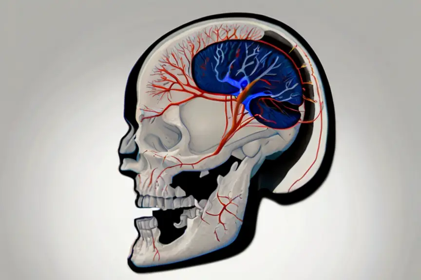
Cerebral angiography is a non-invasive, diagnostic medical procedure that uses contrast dye and X-rays to visualize the blood vessels in the brain.
It is an essential tool in diagnosing and treating various brain vascular conditions, such as blockages, aneurysms, arteriovenous malformations, blood clots, and vasculitis.
In this blog post, we will delve into the world of cerebral angiography, discussing its purpose, common reasons for performing the procedure, and the benefits and risks associated with it.
What is Cerebral Angiography?
Cerebral angiography is a diagnostic procedure where a catheter is used to inject contrast dye into the blood vessels of the brain.
This dye helps in making the blood vessels visible on X-ray images, enabling physicians to assess their structure and blood flow.
Typically conducted in a hospital or radiology center, the procedure usually lasts around an hour.
- Read also: A Comprehensive Guide: Various Types of Cerebral Aneurysms
- Read also: Deciphering Cerebral Infarction: Exploring its Pathophysiology

Common reasons for performing a cerebral angiogram
Diagnosing blockages (atherosclerosis)
Atherosclerosis is a condition characterized by the buildup of plaque in the arteries, which can lead to narrowing and blockages.
Cerebral angiography is often used to diagnose and evaluate the severity of atherosclerosis in the brain, as well as to plan potential treatment options such as angioplasty or bypass surgery.
Identifying aneurysms (weak bulges)
Aneurysms are weak bulges in the blood vessel walls that can rupture and cause bleeding in the brain.
Cerebral angiography is an effective way to identify aneurysms and determine their size, location, and potential risk of rupture.
This information is crucial for planning appropriate treatment, such as surgical clipping or endovascular coiling.
Detecting arteriovenous malformations (abnormal connections)
Arteriovenous malformations (AVMs) are abnormal connections between arteries and veins in the brain that can cause bleeding and other complications.
Cerebral angiography is used to locate and evaluate the size and complexity of AVMs, guiding treatment decisions such as surgical resection or embolization.
Investigating blood clots
Blood clots in the brain can cause stroke or other neurological complications.
Cerebral angiography can help identify the source of these clots, allowing for targeted treatment and potential prevention of future clots.
Evaluating vasculitis (inflammation of blood vessels)
Vasculitis is a condition that causes inflammation and damage to blood vessels, which can lead to various complications.
Cerebral angiography can help diagnose vasculitis and assess the extent of damage, aiding in the development of a treatment plan.

Main steps involved in a cerebral angiogram
The cerebral angiogram procedure involves several key steps to ensure accurate results and patient comfort.
Here’s a detailed breakdown:
Preparation (fasting, medication adjustments)
Before the procedure, patients are advised to fast for several hours to ensure an empty stomach, reducing the risk of complications during the test.
Additionally, medications that might interfere with the procedure or the contrast dye may need to be adjusted or temporarily stopped.
This step is crucial for ensuring the safety and effectiveness of the angiogram.
Catheter insertion (groin or arm artery)
The next step involves the insertion of a thin, flexible catheter into either the groin or the arm artery.
This catheter serves as a conduit for delivering the contrast dye to the blood vessels of the brain.
The choice of insertion site depends on factors such as patient preference, the physician’s expertise, and the patient’s medical history.
Contrast dye injection and X-ray imaging
Once the catheter is in place, the contrast dye is injected through it into the blood vessels of the brain.
This dye is specially formulated to make the blood vessels visible on X-ray images, allowing the physician to obtain detailed images of the brain’s vascular anatomy.
The X-ray machine captures real-time images as the dye flows through the blood vessels, providing valuable information about blood flow patterns and detecting any abnormalities.
Catheter removal and recovery
After the imaging is complete, the catheter is carefully removed, and pressure is applied to the insertion site to prevent bleeding.
Patients are then monitored for a brief period in a recovery area to ensure there are no immediate complications.
It’s common for patients to experience mild discomfort or bruising at the insertion site, but these symptoms typically resolve quickly.

Benefits and Risks of Cerebral Angiography
Cerebral angiography offers several benefits and carries certain risks that patients should be aware of before undergoing the procedure.
Advantages
Detailed and accurate images
One of the primary advantages of cerebral angiography is its ability to provide highly detailed and accurate images of the blood vessels in the brain.
These images help physicians visualize the intricate network of blood vessels and identify any abnormalities or blockages that may be affecting blood flow to the brain.
Combined diagnosis and treatment
Cerebral angiography not only allows for the diagnosis of vascular conditions but also offers the potential for simultaneous treatment during the procedure.
For example, if a blockage or aneurysm is detected during the angiogram, physicians may be able to perform interventions such as balloon angioplasty or stent placement to restore blood flow and prevent further complications.
Risks
Allergic reaction to contrast dye
One of the main risks associated with cerebral angiography is the possibility of an allergic reaction to the contrast dye used during the procedure.
While rare, some patients may experience symptoms such as hives, itching, or difficulty breathing shortly after receiving the dye.
In severe cases, allergic reactions can lead to anaphylaxis, a life-threatening condition that requires immediate medical attention.
Bleeding or infection at the insertion site
Another potential risk of cerebral angiography is bleeding or infection at the site where the catheter is inserted into the artery, typically in the groin or arm.
While uncommon, these complications can occur, particularly in patients with underlying medical conditions that affect blood clotting or immune function.
Stroke
Although rare, cerebral angiography carries a small risk of causing a stroke, particularly in patients with pre-existing vascular disease or other risk factors.
During the procedure, manipulation of the catheter within the blood vessels of the brain can dislodge small blood clots or cause injury to the blood vessel walls, potentially leading to a stroke.

- Read also: Understanding the Impact: Damage to the Cerebral Cortex
- Read also: Cerebral Cortex Vs Cerebrum: The Brain’s Key Components
Conclusion
Cerebral angiography is a valuable diagnostic tool for identifying and treating various brain vascular conditions.
Its benefits include detailed imaging and the potential for combined diagnosis and treatment.
However, like any medical procedure, it carries risks such as allergic reactions and bleeding.
It is essential to discuss these risks with your healthcare provider and weigh them against the potential benefits of the procedure.


