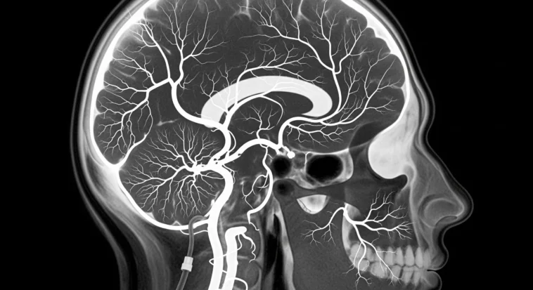
When doctors need the clearest possible look inside the brain, brain angiography is their superhero tool. It lets them spot dangerous problems early—like aneurysms or blocked vessels—before they turn into something serious. If you’ve ever wondered how doctors can actually see blood moving inside your brain, this is how.

What Is Brain Angiography?
Think of brain angiography as Google Maps for your brain’s blood vessels.
Doctors inject a special dye that lights up your arteries and veins on X-rays, showing blood flow in real time. It’s way more detailed than regular scans—like switching from blurry photos to HD video.
I once heard a doctor say it’s like “turning the lights on in a dark room”—suddenly, hidden problems become obvious.
That’s why it’s the gold standard for finding serious brain vessel issues. It’s precise, powerful, and honestly kind of amazing.
- Read Also: A new approach to brain cancer treatment using nano technology has promise
- Read Also: What is neuroplasticity and how does it work? A psychologist explains
Types of Brain Angiography
Here’s the cool part—doctors don’t have just one way to look at brain blood vessels. They’ve got options, like choosing the right tool for a video game level.
Conventional Catheter Angiography
This is the OG, boss-level technique. A tiny tube goes in through the groin and travels up to the brain (yeah, wild).
It’s more intense, but the images are insanely clear—like seeing every crack in the road. Doctors use this when details really matter.
CT Angiography (CTA)
CTA is the fast and efficient option. No catheter threading—just dye and a quick CT scan that builds a 3D picture. It’s perfect in emergencies, when doctors need answers now, not later.
MR Angiography (MRA)
MRA is the gentle genius of the group. No radiation, sometimes no dye, just magnets doing their thing. It’s safer for certain patients, but it takes longer and isn’t always as sharp as the OG method.
Why Doctors Recommend Brain Angiography
Healthcare providers may recommend brain angiography for various diagnostic and therapeutic purposes:
- Detecting Aneurysms: Brain angiography excels at identifying aneurysms, which are weak, bulging areas in blood vessel walls that could rupture and cause life-threatening bleeding.
- Evaluating Stroke Risk: The procedure helps identify blockages, narrowing, or other abnormalities in brain arteries that could lead to ischemic stroke.
- Diagnosing Arteriovenous Malformations: These abnormal tangles of blood vessels can be clearly visualized through angiography, helping doctors plan appropriate treatment.
- Assessing Tumors: Angiography reveals the blood supply to brain tumors, providing crucial information for surgical planning and determining tumor characteristics.
- Planning Interventions: Beyond diagnosis, angiography serves as a roadmap for minimally invasive procedures such as coiling aneurysms or delivering clot-busting medications directly to affected areas.
Research published in medical journals consistently demonstrates that early detection through angiography significantly improves patient outcomes for cerebrovascular conditions.
The Brain Angiography Procedure: What to Expect
Let’s be real, medical procedures sound scary. But knowing what happens step by step makes it way less intimidating.
Before the Procedure
You’ll usually skip food for a few hours (annoying, I know). Doctors will ask about meds, allergies, or kidney issues—basically a safety check so nothing weird happens.
Think of it as prepping your body like a game character before a big mission.
During the Procedure
For conventional angiography, the process unfolds as follows:
- The patient lies on an examination table while monitoring equipment tracks vital signs
- Local anesthesia numbs the catheter insertion site, usually in the groin
- A small incision allows the radiologist to insert a catheter into the femoral artery
- Using fluoroscopy guidance, the catheter is carefully threaded through blood vessels to reach the brain
- Contrast dye is injected while rapid X-ray images capture blood flow patterns
- The radiologist may reposition the catheter to examine different blood vessels
- Once imaging is complete, the catheter is removed and pressure is applied to prevent bleeding
The entire procedure typically takes between one and three hours, depending on complexity. Patients remain conscious but may receive mild sedation for comfort.
After the Procedure
If you had the catheter version, you’ll lie flat for a few hours—perfect excuse to do absolutely nothing. Nurses keep an eye on you, and most people go home the same day.
You might feel sore, get a mild headache, or feel a warm “whoa” sensation from the dye. Serious problems are super rare, and doctors are watching closely the whole time.
Benefits of Brain Angiography
The advantages of brain angiography make it an invaluable diagnostic tool:
- Exceptional Detail: Provides the most comprehensive visualization of brain blood vessels available
- Real-Time Imaging: Captures blood flow dynamics, not just static vessel structure
- Treatment Capability: Can transition seamlessly from diagnosis to minimally invasive treatment
- Accuracy: Offers superior accuracy compared to non-invasive imaging methods for certain conditions
- Versatility: Useful for diagnosing a wide range of cerebrovascular disorders
Medical literature confirms that angiography’s diagnostic accuracy often exceeds 95% for detecting significant vascular abnormalities.
Risks and Considerations
Okay, real talk—no medical procedure is totally risk-free, but brain angiography is very safe. Most people just get a little bruise or soreness where the catheter went in.
Serious stuff like stroke or allergic reactions? Super rare—less than 2%, especially when experts do it.
There’s some radiation, but modern machines keep it low. And if someone’s pregnant or has kidney issues, doctors plan extra carefully or choose another option.
Advances in Brain Angiography Technology
Medical technology continues to enhance brain angiography capabilities:
- 3D Rotational Angiography: Advanced software reconstructs images into three-dimensional models, providing unprecedented views of complex vascular structures.
- Cone-Beam CT: Combines the benefits of CT scanning with traditional angiography, offering detailed images during interventional procedures.
- Artificial Intelligence Integration: AI algorithms now assist radiologists in detecting subtle abnormalities and predicting treatment outcomes.
- Reduced Radiation Protocols: Newer imaging equipment delivers diagnostic-quality images with significantly less radiation exposure.
These technological improvements have made brain angiography safer, faster, and more accurate than ever before.
Alternative Imaging Options
Doctors don’t jump straight to angiography for everyone. CT scans and MRIs are like the “first level” scans—fast and non-invasive.
Ultrasound can even check blood flow in the neck without needles at all. Angiography comes in when doctors need the ultimate close-up.
Making an Informed Decision
If your doctor recommends brain angiography, consider asking these important questions:
- Why is angiography necessary in my specific case?
- Are there alternative imaging options that might provide the information needed?
- What are the specific risks based on my health history?
- How experienced is the interventional radiologist performing the procedure?
- What happens if the angiography reveals an abnormality requiring treatment?
- What is the expected recovery time and what restrictions will I face afterward?
Open communication with your healthcare team ensures you understand both the necessity of the procedure and what to expect throughout the process.
- Read Also: Scientist Revealed – Human Brains Are Getting Smaller Over Time
- Read Also: Why Does Brain Freeze Happen – Neuroscientists Explains
Conclusion
Brain angiography is like turning on stadium lights inside the brain—suddenly, everything becomes clear. It helps doctors catch dangerous problems early and plan life-saving treatments.
Yes, it sounds intense. But millions of people go through it safely every year.
If your doctor recommends it, it’s because the answers it gives are powerful—and those answers help protect something pretty important: your brain.